Anatomy of the Ankle

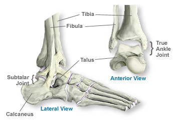
The ankle is a complex mechanism. What we normally think of as the ankle is actually made up of two joints: the subtalar joint and the true ankle joint. The true ankle joint is composed of three bones, seen above from a front, or anterior, view: the tibia which forms the inside, or medial, portion of the ankle; the fibula which forms the lateral, or outside portion of the ankle; and the talus underneath. The true ankle joint is responsible for the up-and-down motion of the foot.
Beneath the true ankle joint is the second part of the ankle, the subtalar joint, which consists of the talus on top and calcaneus on the bottom. The subtalar joint allows side-to-side motion of the foot.

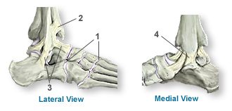
The ends of the bones in these joints are covered by articular cartilage (1). The major ligaments of the ankle are: the anterior tibiofibular ligament (2), which connects the tibia to the fibula; the lateral collateral ligaments (3), which attach the fibula to the calcaneus and gives the ankle lateral stability; and, on the medial side of the ankle, the deltoid ligaments (4), which connect the tibia to the talus and calcaneus and provide medial stability.
These components of your ankle, along with the muscles and tendons of your lower leg, work together to handle the stress your ankle endures as you walk, run, and jump.

